


Transforming Cellular Imaging with Advanced Nanobody innovations
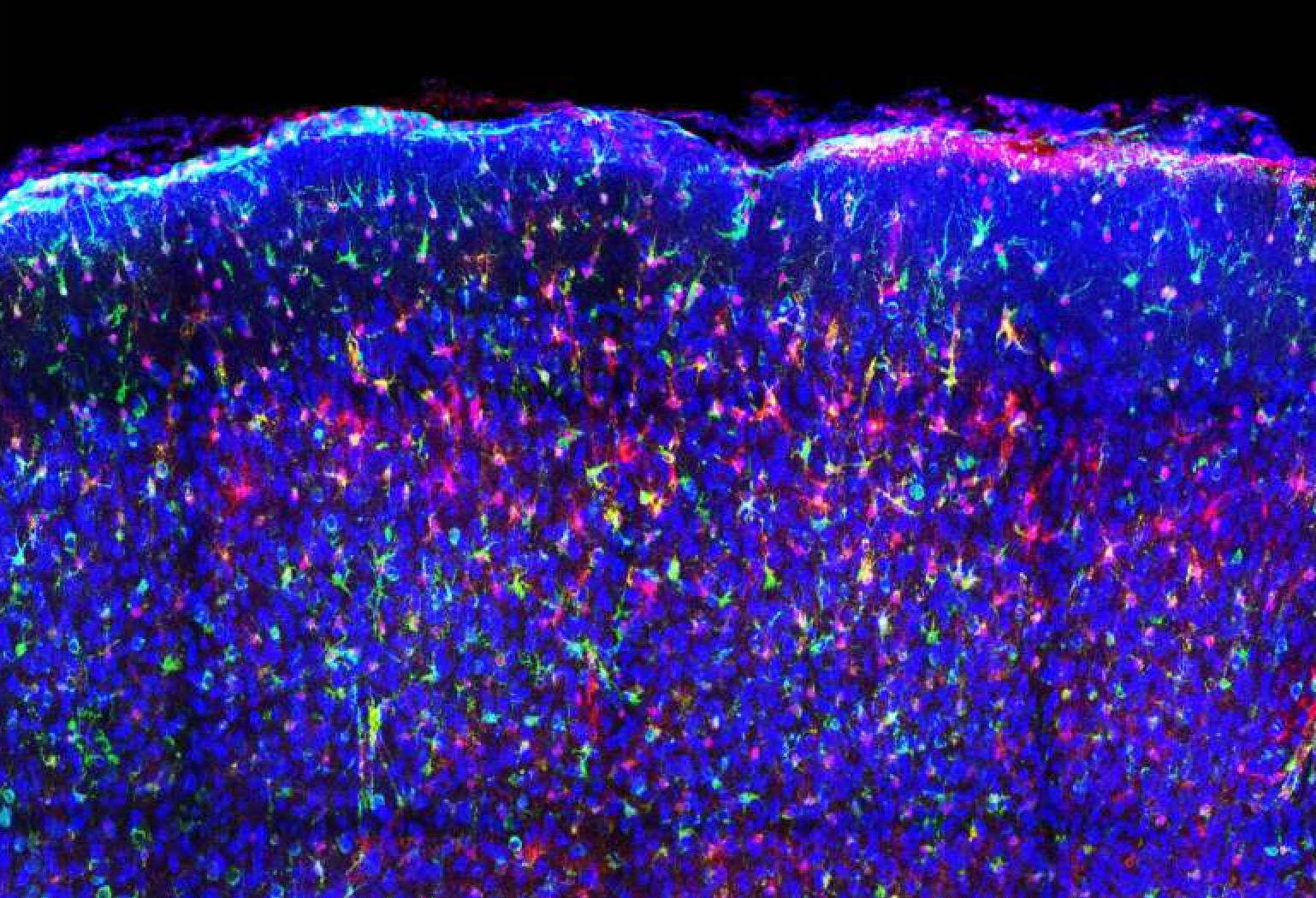
Vibrant multicolor depiction of mouse brain tissue revealing astrocytes in purple alongside neurons in blue, exemplifying precise cell-type-specific visualization.
Limitations of Customary Antibodies Within Cells
For decades, antibodies have been pivotal tools in medicine for neutralizing pathogens, identifying malignant cells, and diagnosing illnesses. However, their functionality is predominantly restricted to extracellular spaces. The intricate surroundings inside cells presents a formidable challenge because conventional antibodies are generally too large and delicate to function effectively within the intracellular landscape where many diseases originate.
This constraint arises as standard antibodies evolved to operate mainly within bodily fluids such as blood serum. When introduced into the cellular interior, they often lose their structural stability or clump together, which compromises their effectiveness. Even fluorescently tagged versions frequently suffer from instability or diminished binding capacity when used inside living cells.
Nanobodies: Compact Molecular Probes for Intracellular Targeting
A revolutionary solution has emerged through nanobodies-small antibody fragments derived from camelids like llamas and alpacas-that can infiltrate living cells while maintaining robust stability bound to specific intracellular molecules.These nanobodies are engineered by embedding fluorescent tags within flexible internal regions rather then at terminal ends, preserving both brightness and target affinity.
This design innovation yields a spectrum of durable probes emitting vivid colors including blue, green, orange, red, and near-infrared wavelengths. Each probe selectively attaches to proteins ranging from ubiquitous cellular markers to viral elements or structural components involved in intercellular dialogue pathways.
Enhanced Fluorescence specificity Reduces Background Interference
The fluorescence signal activates only upon direct interaction with the intended target inside the cell’s environment. This selective activation significantly diminishes background noise that typically obscures imaging clarity during live-cell microscopy sessions.
Diverse Applications Demonstrating Real-Time Cellular Dynamics
- Cultured human cells: Distinct organelles such as nuclei membranes and mitochondria were vividly illuminated without affecting cell health or function.
- Neuronal signaling: Calcium ion fluxes-a key neural communication mechanism-were tracked dynamically to reveal active brain processes with high temporal resolution.
- In vivo mouse models: Deep-brain imaging captured targeted neuronal populations exhibiting sustained fluorescence suitable for cutting-edge neuroimaging techniques like two-photon microscopy.
- zebrafish embryogenesis: Multicolor labeling enabled simultaneous observation of developmental signaling cascades during early growth phases providing insights into complex morphogenetic events.
The Advantage of Multiplexed Intracellular Visualization
A major leap beyond traditional approaches is this technology’s ability for multiplexed imaging-together monitoring multiple molecular interactions using distinct fluorescent colors within single live cells. While conventional methods often limit researchers to one or two color channels, multicolor imaging enables comprehensive mapping of intricate protein networks implicated in multifactorial diseases such as cancer metastasis or neurodegenerative disorders where numerous pathways converge simultaneously.
A Comprehensive Outlook on Cellular Pathology
This expanded color palette grants scientists unparalleled insight into how diverse cellular components coordinate during disease progression by capturing multiple biological events concurrently instead of isolated snapshots alone.
Navigating Delivery Challenges Toward In Vivo Implementation
An ongoing obstacle remains developing efficient delivery systems capable of introducing these fluorescent nanobody probes directly into tissues under physiological conditions.Their immediate impact is nonetheless profound: researchers can now visualize intact intracellular architectures dynamically without resorting to invasive sample processing that disrupts native states.
This breakthrough represents a paradigm shift by converting fluorescent antibody fragments into real-time molecular beacons illuminating previously hidden inner workings within living organisms across various biological contexts.

